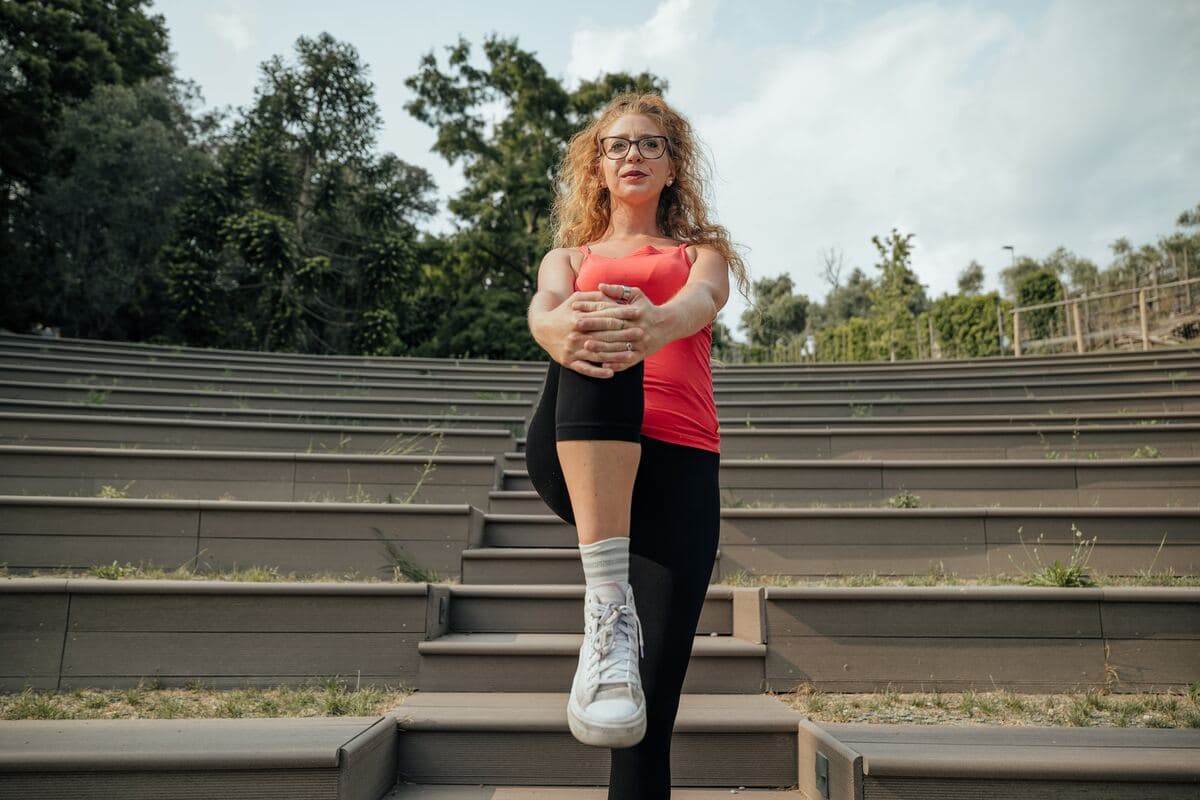
The knees touch, the ankles stay apart, and in between an X forms. This is knock knee (genu valgum), and on the internet it is almost always presented as a countdown: arthritis, pain, ligaments at risk, "correct it while you still can". The research tells a calmer and more useful story, and it is the story worth knowing before starting any programme.
In short: in the great majority of cases knock knee is a variation in shape without symptoms or limitations, valgus alignment on its own does not start arthritis in a healthy knee, and no exercise straightens a bone. This does not mean there is nothing to do: it means exercises should be judged on what they really change: movement control, pain, the risk of injury. Below you will find what is documented, what is not, and how to use this information anyway.
⚠ Important: read this before continuing
The exercises and advice in this article are general indications and may not suit your situation. Every person has a unique clinical history: what works for others could be counterproductive for you.
Want a professional's opinion? Start with the questionnaire: you register and see who will follow you, a physiotherapist registered with the Register chosen for your case, whom you can write to in chat. If you would rather hear a voice, the first consultation is free and we look at your situation together.
First of all: when knock knees need checking, not training
Knock knee in itself is not an emergency. Some situations can present alongside it, and they need to be assessed before starting any exercise:
- One knee in valgus, while the other is straight. Asymmetric valgus is not the common variation in shape: it can arise from a previous injury, damage to the growth plate in childhood, a joint condition or a bone disease. It needs investigating, not training.
- A deformity that appears or worsens quickly in an adult. The alignment of the legs does not change from one month to the next for no reason.
- A hot, swollen and red knee, perhaps with fever or general malaise: this can be acute inflammation or a joint infection, and it is a situation to assess promptly. We cover it in detail in the article on a hot, swollen knee.
- Swelling that appeared within hours after an injury, with the feeling that the knee "doesn't hold".
- Joint locking or sudden giving way while walking, even without pain beforehand.
- Pain that wakes you at night or that never changes, in any position, whatever you do.
- In children: valgus that worsens after age 7 instead of reducing, very marked valgus, valgus on one side only, short stature or associated pain.
None of these pictures improves by doing clamshells. Time spent strengthening the gluteus medius in these cases is time taken away from the diagnosis.
What everyone calls it
On the medical report and from the orthopaedic surgeon you will read valgus or genu valgum. In everyday speech there are two other expressions, and this is how most people search for it: knock knees and X legs, because seen from the front, with the knees touching and the ankles apart, the legs form exactly that letter. It is the same thing. The opposite, with the knees apart and the ankles close together, is called bow legs or genu varum, or O legs.

Knock knee: what it is, and the distinction that changes everything
Standing upright with the knees in contact, the inner ankle bones normally touch or stay very close together. In valgus the knees touch and the ankles do not. The clinical measure used to quantify it is the intermalleolar distance: you stand with the femoral condyles in contact and measure how far apart the ankles are. A distance above 8 cm is considered pathological as a reference threshold, which is a different thing from a diagnosis, and very different from a prognosis.
Then there is the Q angle, the angle between the axis of the femur and that of the patellar tendon, which for years was the iconic number of valgus. On this, research has been clear, and it needs saying straight away because it shapes half of the article you are reading: in a systematic review of prospective studies on almost 5,000 people, the Q angle was not found to be a risk factor for future patellofemoral pain. Measuring it does not tell you who will have pain.
The genuinely useful distinction is another one, and it is the one that common talk constantly confuses:
Structural (static) valgus: this is the shape of the bones and the orientation of the joint surfaces. It is seen standing still. It does not change with training, because no muscle contraction alters the geometry of a femur. In adolescence a minimal or no change in alignment is now expected.
Dynamic valgus: this is the knee collapsing inwards during a movement, such as a squat, a step down the stairs or a landing from a jump. It is a motor behaviour, not a shape, and like all behaviours it can be trained.
When you read "exercises to correct knock knees", in almost every case the second is being discussed. Confusing the two means promising something that cannot happen and then making those who "did not manage it" feel guilty.
Valgus can also be bilateral, the far more common form, often simply the conformation a person is built with, or unilateral, which as mentioned above deserves investigation because it is more often linked to a specific cause.
The causes of knock knee, put into proportion
Physiological development. In children valgus appears around age 2, is most noticeable between 3 and 4, and then reduces to a mild, stable valgus by around age 7. In many adults what remains is simply the point at which that process stopped. The same applies in the rotational plane: feet turned in or out also have an age at which they appear and one at which they resolve, which is why they are not treated: the full picture is in children who walk with their feet turned in or out.
Weakness of the gluteus medius and hip external rotators. This is the explanation most repeated in gyms and physiotherapy studies, and it is the one that holds up least well. A systematic review with partial meta-analysis gathered 22 studies that measured hip strength and dynamic valgus in healthy people: for the abductors, 12 studies found no significant correlation, 2 supported it and 2 went in the opposite direction; for the external rotators, 8 found no correlation, 2 were in favour and 2 against. None of the partial meta-analyses reached statistical significance. The authors' conclusion is that hip weakness may not be the cause of dynamic valgus.
The same holds in the direction of pain: the review of prospective studies on patellofemoral pain found that hip weakness did not predict who would develop pain in future, and that in adolescents it was, if anything, greater hip abduction strength that was associated with risk. In the same review, however, one predictive factor did emerge: quadriceps weakness in military recruits, especially when normalised to body mass index. It is not a detail to hide in order to make the argument fit: it is a clue about where it is worth looking.
A tight iliotibial band. It appears in almost every article on the subject as a mechanical cause of valgus. We did not find literature supporting it, and the previous version of this article did contain this claim: we have removed it. Foam rolling the outer thigh can feel pleasant and temporarily reduce local sensitivity, but it should not be sold as a correction of alignment.
Flat feet and overpronation. The idea that the foot's contact with the ground drags the tibia into rotation and "creates" valgus is plausible in theory, and in practice it holds up less well than expected: a randomised study of people with patellofemoral pain stratified participants precisely by midfoot mobility and found that this did not allow prediction of who would respond better to orthoses than to hip exercises. The foot matters, but not in the linear way it is often described.
Trauma, fractures and bone or metabolic causes. These are the causes of genuine structural valgus, particularly asymmetric cases or those that worsen during growth. Rickets and other metabolic or genetic conditions belong here and must be ruled out by a specialist, not managed with an exercise programme.
Body weight. Excess weight is often cited among the causes. We do not have solid evidence that losing weight changes alignment; what matters is a different question, namely how much load crosses a joint that is already symptomatic. We discuss it in body weight and the knee.
An important tip: before trying these exercises, make sure you have had a correct diagnosis. People often think they have one problem, but the real cause of the pain is something else. Doing the wrong exercises can make things worse rather than better.
Knock knee: real consequences and presumed ones
Arthritis: the precise data
Here the literature is clearer than it seems, and it says two different things depending on where you start. A longitudinal study of a large cohort measured alignment with full-limb radiographs and followed the knees for 30 months.
In knees without arthritis at the start (almost 3,000), alignment in varus increased the risk of developing arthritis; alignment in valgus did not. In knees that already had arthritis, on the other hand, valgus almost quintupled the risk of progression in the lateral compartment, and reduced that of medial progression.
Translated: if you have X legs and the cartilage is healthy, valgus alone is not the spark that lights arthritis. If arthritis is already present, alignment matters a great deal for where and how quickly it worsens, and the part under pressure is the outer one, not the inner. This is the exact opposite of what is often read, and of what this very article said before it was rewritten. If the topic concerns you, we cover the full picture in knee osteoarthritis.
Patellofemoral pain
The association between valgus and anterior knee pain exists in clinical observations, but as a predictive factor it did not hold up: neither the Q angle nor hip strength anticipated who would develop the problem. This does not remove patellofemoral pain from the picture, since it is one of the most common conditions of all, but it shifts the reasoning from the shape of the leg to how the leg is loaded. If that is your case, the pathway is described in patellofemoral syndrome.
Anterior cruciate ligament
This is the most instructive story of all. A 2005 prospective study of 205 female athletes, of whom only 9 later tore their cruciate ligament, had found that the knee's abduction angle and moment on landing predicted the injury, and it is from there that the entire industry of dynamic valgus screening was born. Nine cases is a very small number, and the authors of the later verification say so explicitly: the hypothesis rested on a single, small sample. In 2016 the verification came in a study of 710 elite Norwegian female football and handball players followed for up to 42 new non-contact injuries: among the five variables tested, only medial displacement of the knee was associated with risk, but with an area under the curve of 0.6, meaning a test to be discarded for its ability to discriminate. And among the 643 athletes with no previous injury, no jump variable was associated with risk. The factor that really carried weight was having already had a cruciate injury.
It is therefore not possible to watch someone descend into a squat, see the knee collapse inwards and tell them they will tear their cruciate. You can explore the rest of the picture in cruciate ligament rehabilitation.
So why train?
Because the failure of screening is not the failure of training, and this is the constructive part. A meta-analysis that pooled eight meta-analyses on injury prevention programmes for the cruciate found a 50% reduction in cruciate injury risk across all athletes, and 67% for non-contact injuries in female athletes. The same authors note that data on males is not sufficient to draw conclusions.
These are neuromuscular training programmes, covering strength, jumps and landings, balance and movement control: the same family of work to which the exercises below belong, with one difference to keep in mind. In the studies these were complete, structured programmes, not a handful of exercises done now and then. They work. They simply do not work because they "straighten" something, and they are not used to select who to train: it is better to train everyone.
Exercises for knock knee: what they can and cannot do
The honest promise is this: these exercises will not change the shape of your legs. They can make the knee stronger and more stable under load, improve movement control in situations where it collapses inwards, reduce pain when it is present, and, in a sporting context, lower the risk of injury. That is not a small thing: it is almost everything that is really needed.
You will not find figures for loads and repetitions in this article, and that is deliberate: the dose that works depends on your starting level, and the fixed guidelines that circulate do not come from any study. The practical criterion you can use is quality: you continue as long as the movement stays clean and the knee does not collapse inwards, and you stop when alignment begins to be lost, not when the count runs out.
Hip strength
These remain in the programme, not as a "correction", but because a strong leg handles load better.
Clamshell. Lying on your side, knees bent and feet together: open the knees without rolling the pelvis backwards. If the pelvis opens, the exercise has gone somewhere else.
Side-lying leg raise. On your side, upper leg straight lifting while staying in line with the body, without bringing it forward.
Monster walk. A band above the knees, a sideways walk in a half squat keeping constant tension on the band and without straightening the legs with each step.
Fire hydrant. On all fours, bring the knee out to the side keeping the trunk still and without tilting the pelvis to gain range.
Bridge with abduction. During the bridge, press the knees outwards against a band while keeping the pelvis high and stable.
External rotation with band. Seated, with a band around the knees, open the knees against resistance while also controlling the return.
Quadriceps
This is the only group that, in the review of prospective studies, emerged as a risk factor when weak, and it needs stating precisely: the finding concerns military recruits, not the general population. It is nonetheless the group most often neglected in "anti-valgus" programmes in favour of the glutes. Squats, leg press, step-ups, progressively loaded lunges and open chain work all have their place. If you are starting from zero, a reasoned starting point is in knee exercises.
Movement control
Here the work is on dynamic valgus, that is, the part of the picture that depends on how you move and not on how you are built: it is the only part training can influence. The principle is always the same: a task difficult enough to challenge you, but not so difficult that you lose alignment.
Squat with a band above the knees, pushing against the resistance to keep the knees over the line of the feet.
Single-leg squat, checking that the knee does not collapse inwards and that the pelvis does not drop on the free side.
Step down. Standing on a step, lower yourself slowly on one leg: this is the movement in which collapse is most visible, and the one that most resembles the stairs at home.
Lunges and walking lunges, keeping the knee aligned over the foot throughout the descent.
Landings. In sport, jumping and landing in a controlled way is the part of neuromuscular work that weighs most heavily in prevention programmes. It should be introduced gradually and once strength is in place.
Mobility and soft tissues
Stretching the adductors and working on the outer side of the thigh can make sense for how they make you feel and for your tolerance of movement. We no longer present them as corrections of alignment, because they are not.
The programme, exercise by exercise
What follows is the work that makes sense to do: not to straighten the bone, but to build the hip control that governs how the knee behaves under load. Two or three times a week, with progression.
Clamshell
It serves to activate the glutes in an unloaded position, without body weight forcing you to compensate. It is the classic starting point when the hip struggles to take part in the movement.
Lie on your side with the knees bent to 90 degrees and the feet together. Keeping the feet in contact, lift the upper knee, opening the legs like a shell. The movement should be slow and controlled, without rolling the pelvis backwards to gain range.
A practical starting point is 15 to 20 repetitions per side, three sets. To make the exercise more demanding, add a band above the knees. You should feel the glute working, not the outer thigh or the lower back.
Monster walk
Compared with the clamshell, here you are weight-bearing, and the task becomes maintaining the position of the hip while you move. It is much closer to what you ask of your body in real life.
Place a band above the knees or around the ankles. Bend slightly into a mid-squat and walk sideways, keeping the band under constant tension. The pelvis stays stable: if it swings with every step, the band is too stiff.
Ten to fifteen steps per direction, three sets. The knees stay aligned with the toes throughout, and the torso does not lean towards the side it is moving to.
Single-leg glute bridge
This is the first genuinely demanding exercise on the list, because it removes a support and forces the pelvis to stabilise by itself.
Lie on your back with one knee bent and the foot on the floor, the other leg extended in the air. Lift the pelvis by pressing through the heel of the supporting foot and contracting the glute. The two iliac crests must stay at the same height: if one side drops, you have exceeded your current level.
Ten to twelve repetitions per side, three sets. If the pelvis rotates or sinks, reduce the range of the movement or temporarily return to the two-leg version. A small, clean movement is better than a large, sloppy one.
Wall sit with a ball
This exercise is isometric: it does not teach a movement, it teaches you to stay under load while holding a position you can recognise.
Lean your back against the wall in a squat position, with the thighs descending towards parallel. Place a soft ball between the knees and squeeze it lightly, activating the adductors. Check that the knees stay above the line of the feet.
Hold the position for 30 to 60 seconds, repeating three or four times. The aim is not to resist for as long as possible, but to stay in the correct position for the whole time you have set: when quality drops, the set is over.
Squat with a band
Here the work moves from muscle to movement. The band does not "straighten" anything: it gives you a tactile reference that makes clear where your knees are, something almost nobody can tell with their eyes closed.
Place a band above the knees. During the descent actively push the knees outwards against the resistance. Descend only as far as you can maintain alignment: that is your useful range today, and it will grow.
Twelve to fifteen repetitions, three sets. It is worth filming yourself from the front from time to time: what you feel your knees are doing and what they actually do almost never match.
Lateral step down
This is the exercise closest to what happens when you go down stairs, land from a jump or run, and for that reason it is also the most selective.
Standing on a step, slowly lower one leg towards the floor by bending the knee of the supporting leg, then come back up. The working knee stays aligned and does not collapse inwards; the pelvis does not sag on the free side.
Eight to ten repetitions per side, three sets. If the knee gives way or the pelvis drops, lower the step: the rule is always the same, you progress in height only when control holds. Single-leg exercises are the most valuable because they expose asymmetries between one side and the other, which movements on two legs keep hidden.
Complementary mobility and stretching
Strengthening alone leaves out a piece. If a joint above or below does not move, the knee ends up compensating.
Shortened hip flexors, typical of people who spend many hours seated, limit extension and change how the pelvis positions itself under load. Calf stretching and work on the lateral tissues of the thigh also have their use as a complement to strengthening.
Ankle mobility is the most overlooked part of all. An ankle that does not dorsiflex well prevents the tibia from advancing during a squat or landing, and the body finds the route left to it, often sideways, through the knee. Before insisting only on the glutes, it is worth testing dorsiflexion and, if needed, adding dedicated ankle mobility work.
How to organise the week
Among the knock knee exercises seen above, the low-demand activation ones (clamshell and monster walk) lend themselves to being done almost every day, including as a warm-up before running or training. They do not fatigue you and prepare the hip to take part.
The more demanding work (single-leg bridge, step down, squat) should be spread over about three sessions a week, leaving at least one rest day between them. Mobility can be daily.
On timelines for results I want to be precise, because this is where people most often mislead: I do not have a number to give you. Studies that measured a change in landing mechanics used interventions of at least four weeks, but that figure describes the length of the protocols, not the moment you will feel better. Take it as the minimum order of magnitude within which it makes sense to assess, not as a promise.
Knock knee: exercises to avoid
Here too the common answer, that squats and lunges should be avoided with X legs, is wrong, for the reason given above: the problem is not the movement, it is control of the movement. Removing squats from someone whose knee collapses inwards leaves them exactly as they were, only weaker.
What is worth avoiding is more precise and shorter:
- Loading heavily while the knee collapses inwards. It is not the squat that is the problem, it is the squat done with the knee caving in under load: there you add weight to a pattern that cannot hold. Reduce the load, recover control, then build back up.
- Impact exercises and changes of direction, if jumps and landings still show collapse: this is the movement in which dynamic valgus is most visible, and it should be built afterwards, not before.
- The adductor machine used to "close" the legs. It is the exercise most requested by people with X legs and one of the least useful: it strengthens the group that draws the thigh towards the inside, which is the direction in which the knee is already collapsing.
- Pushing through a knee that hurts during the exercise. Mild discomfort that passes during the day is fine; pain that increases session after session is a signal, not a milestone.
Knock knee: insoles, taping, weight and surgery
Insoles. International consensus statements on patellofemoral pain recommend orthoses as an option to improve pain and function, and this holds regardless of the shape of your foot: the randomised study comparing orthoses with hip exercises found no difference in success rates (48% against 50%), and above all midfoot mobility did not help to work out who would respond better to what. They are a reasonable option to try, not a correction of the axis.
Taping. The same consensus document places taping and patellar braces among the interventions for which the evidence is uncertain. It is not forbidden to try them, but they are not the lever on which to build the pathway.
Body weight. There is no evidence that losing weight changes alignment; what changes is the load that crosses the joint. In a knee that is already arthritic it is a variable that matters.
Surgery. For marked, symptomatic structural valgus, the option is an osteotomy that realigns the bone and offloads the lateral compartment. It is an orthopaedic decision, reserved for selected cases, and not the "serious" alternative to exercises: it is a different thing, for a different kind of problem.
Knock knees in children
This is the chapter where common sense counts for more than any protocol. Valgus appears around age 2, peaks between 3 and 4, and usually reduces spontaneously to a mild, stable valgus by around age 7. Most children with X legs are asymptomatic and have no functional limitations. Standard management in this age group is observation. Before that stage there is another that worries parents even more, bowed legs in the first year: their peak falls around six months, and it does not depend on standing the baby up.
Specialist assessment is needed when the picture moves off track: valgus that worsens after age 7, very marked, on one side only, associated with pain, with short stature or with a gait with the feet turned outwards. In these cases bone or metabolic causes are sought and, when indicated, residual growth is addressed: decisions that belong to the paediatric orthopaedic surgeon.
What is not needed: "preventive" insoles, sporting bans and corrective programmes for a child who has no symptoms.
How to use this information anyway
Putting things into proportion is not an invitation to do nothing. It is a change of target, and in practice it works like this.
If you have no pain, knock knee is not a problem to solve. Train for the reasons everyone trains, and if you do sports involving jumping and changes of direction, include a complete neuromuscular programme: those work genuinely well.
If you have pain, knock knee is not the diagnosis: it is a detail of appearance. The right question is which structure hurts, in which movements and under which load. For patellofemoral pain, international consensus recommends exercise therapy, in particular the combination of work on the hip and the knee, while advising against isolated mobilisations and physical therapy modalities used on their own.
If you already have arthritis, alignment is genuine clinical information, which guides expectations and decisions about load. It is worth discussing with whoever is following you.
Judge your programme by the right outcomes. Not by a photo of your legs in the mirror after three months: that will not change. By how well you manage stairs, how long pain lasts after a session, how stable you are on one leg. If after several weeks of serious work none of this moves, the problem is not that you did not push hard enough: it is that the hypothesis needs revisiting.
In summary
Knock knee is, in the great majority of cases, a variation in shape, not a disease in incubation. Valgus alignment did not increase the risk of developing arthritis in healthy knees, while it did increase lateral progression in knees already affected by arthritis; the Q angle and hip strength do not predict patellofemoral pain; weakness of the glutes is not shown to be related to dynamic valgus in healthy people; and observing knee collapse during a jump does not allow cruciate injuries to be predicted. At the same time, neuromuscular training programmes halve the risk of cruciate injury, and combined hip-and-knee exercise is recommended for anterior knee pain. The exercises remain; what they promise has changed.
If you have X legs and want to understand whether they really have anything to do with your pain, or simply to build a sensible programme without chasing a correction that will not come, the physiotherapists at Ri-Hub can assess your case through telerehabilitation and set up the work together with you.
You might also be interested in:
Cervical Disc Herniation: Symptoms, Causes and Exercises Preventing Falls in Older Adults: Exercises and Strategies to Reduce Risk Rotator Cuff Exercises: Strengthening, Stretching and Complete Rehabilitation
Stay in touch with us Join our dedicated Facebook group: we answer all patients, give practical advice and clear up the most common questions.
Sources
- Varus and valgus alignment and incident and progressive knee osteoarthritis, Annals of the Rheumatic Diseases, 2010
- Risk factors for patellofemoral pain: a systematic review and meta-analysis, British Journal of Sports Medicine, 2019
- The effect of hip abductor or external rotator strength on dynamic knee valgus in healthy subjects: a systematic review with partial meta-analysis, The Knee, 2026
- The Vertical Drop Jump Is a Poor Screening Test for ACL Injuries in Female Elite Soccer and Handball Players: A Prospective Cohort Study of 710 Athletes, The American Journal of Sports Medicine, 2016
- Meta-analysis of meta-analyses of anterior cruciate ligament injury reduction training programs, Journal of Orthopaedic Research, 2018
- 2018 Consensus statement on exercise therapy and physical interventions (orthoses, taping and manual therapy) to treat patellofemoral pain, British Journal of Sports Medicine, 2018
- Does foot mobility affect the outcome in the management of patellofemoral pain with foot orthoses versus hip exercises? A randomised clinical trial, British Journal of Sports Medicine, 2020
- Biomechanical measures of neuromuscular control and valgus loading of the knee predict anterior cruciate ligament injury risk in female athletes: a prospective study, The American Journal of Sports Medicine, 2005
- Genu Valgum, StatPearls, updated 2023
Last source check: August 2026.
Need a consultation?
Start with the questionnaire on Ri-Hub: a few questions about your case, then you register and see who will follow you, a physiotherapist registered with the Register, whom you can write to in chat whenever you like. If you would rather talk it through by voice, the first consultation is free, lasts twenty minutes and commits you to nothing: it starts with them already having read what you wrote, and together you decide whether and how to go on.