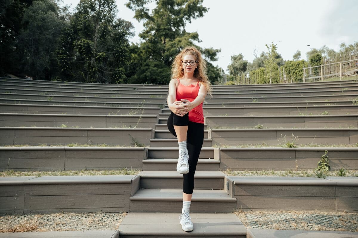


Hip pain during squats: it is one of the most frequent reasons people who train stop going down, change exercise or abandon the barbell bench altogether. The instinctive reaction is to look for the technical error: foot width, depth, the pelvis rolling under at the bottom. This search has far less foundation than the web makes it seem.
This article sets out what is really known: which structures can hurt, why watching the movement is not enough to tell which one it is, what studies have shown about technical changes and what, instead, has evidence of working. And, first of all, when hip pain during squats is not a question of training but a reason to stop.
⚠ Important: read this before continuing
The exercises and advice in this article are general indications and may not suit your situation. Every person has a unique clinical history: what works for others could be counterproductive for you.
Want a professional's opinion? Start with the questionnaire: you register and see who will follow you, a physiotherapist registered with the Register chosen for your case, to whom you write in chat. If you would rather hear their voice, the first consultation is free and we look at your situation together.
First of all: when it is not a technique problem
There are situations in which changing your squat set-up is the wrong thing to do, because the problem is not the movement.
The most important is the stress fracture of the femoral neck. In the review published in the Journal of the American Academy of Orthopaedic Surgeons, the typical picture is hip and groin pain with a gradual onset, no trauma, that eases with rest; it mainly affects military recruits and endurance athletes. The point that makes this condition insidious is another: X-rays are often normal, and it is magnetic resonance imaging that has clearly superior sensitivity and specificity. If it is not recognised, the lesion can progress to a complete, displaced fracture, with a risk of non-union and necrosis of the femoral head.
One point is almost always missed: the fact that the pain goes away when you stand still rules nothing out. In the classic description of femoral neck stress injury, rest eases the pain exactly as you would expect from a "muscular" problem, and this is one of the reasons the diagnosis arrives late. Stop and get assessed, rather than looking for corrections, if one of these circumstances applies:
- groin pain that appeared with no specific episode and continues even when walking, running or climbing stairs, even if it settles at rest;
- an important and recent increase in training volume, running or load;
- pain that wakes you at night or does not ease in any position;
- pain accompanied by fever, general malaise or unexplained weight loss;
- sudden giving way of the limb, a feeling of a true joint block, or pain after a fall or a trauma;
- in the growing adolescent, hip or knee pain with a limp: a problem with the growth plate needs to be ruled out.
The first two points are the picture described by the review on stress injury; the others are general reasons for caution that apply to any musculoskeletal pain. None of these points is resolved by widening your foot stance. All of them are resolved by an assessment.
What research really says (the uncomfortable part)
Here comes the part that almost no article on the topic tells, and it completely changes how the problem should be approached.
There is no recognisable "squat of a person with a painful hip" that you can spot by eye. A study published in Medicine & Science in Sports & Exercise compared 15 people with symptomatic femoroacetabular impingement, already scheduled for surgery, with 14 healthy controls of the same age and sex, using three-dimensional movement analysis. The result: no difference in squat depth, in either of the tasks tested. The differences that emerged were others, and much less visible: in the constrained version of the exercise, greater rise of the pelvis and more hip adduction on the symptomatic side; in the free squat, a slower descent; and on the way down, a smaller external rotation moment at the hip, with less variability of the angle in the transverse plane. These are quantities measured in the laboratory, not things you see by watching someone squat.
The systematic review with meta-analysis in the British Journal of Sports Medicine, which gathered 14 studies of low to moderate quality, reaches a similar conclusion, cautiously: people with the syndrome squat to a lesser depth but without differences in hip range of motion in flexion, and the authors stress that the differences between groups are small to moderate. In plain terms: these are laboratory averages, not a fault you can recognise in the gym.
And indeed, when someone tried to recognise it in the gym, they could not. A study in the Journal of Sport Rehabilitation filmed the single-leg squat test in a clinic, on 20 men with bilateral femoroacetabular impingement and 20 asymptomatic men, measuring depth, pelvic drop, hip adduction and knee valgus: none of the measures distinguished the two groups.
There is a second finding that dismantles the most common explanation. The systematic review of the radiological prevalence of femoroacetabular impingement, published in the European Journal of Radiology across 60 studies, concludes that imaging signs attributed to impingement are common in athletes, in asymptomatic people and in symptomatic patients. They are indeed more frequent in symptomatic patients, and the mean alpha angle is greater in those with symptoms, but the same bone shape is found in a great many people who feel absolutely nothing. It is no accident that the Warwick international consensus defines femoroacetabular impingement syndrome as the combination of symptoms, clinical signs and imaging findings: a report alone is not a diagnosis.
What do we take from this, in practical terms? That hip pain during squats is not resolved by finding "the error". It is addressed by understanding which structure is irritated, how much load it tolerates today, and how to bring it gradually back to tolerating more. Technical changes remain very useful, but as tools for continuing to train, not as cures.

Where it hurts: the map that really matters
Location does not make a diagnosis, but it guides the hypotheses and helps you describe the problem clearly to whoever assesses you.
Groin and the front fold of the hip, especially at the bottom of the descent. This is the most typical site of an intra-articular problem: labrum, capsule, cartilage, or the impingement syndrome. Often the patient points to the spot by pinching the thumb and index finger just above the fold: the classic "C sign".
Side of the hip, above the greater trochanter. Here the most frequent suspicion is gluteal tendinopathy, which causes pain even lying on that side and when climbing stairs. It is worth noting that the most robust clinical study on this picture, the one we discuss further on, enrolled participants with a diagnosis of gluteal tendinopathy confirmed on MRI, not of "bursitis": it is the tendon that is the target of treatment. We discuss it in detail in the article on lateral hip pain.
Inner groin, towards the inside of the thigh. It points to the adductors, especially if the pain is reproduced by squeezing your knees together against resistance.
Deep or posterior gluteal pain. Here the problem can also start elsewhere, for example in the lumbar spine: if the picture is unclear, the overview of the causes of hip pain helps you put things in order.
One detail matters more than location: how the pain behaves. Does it appear only beyond a certain depth or also bodyweight? Does it warm up and improve after the first sets, or worsen set after set? Does it come back the next morning? These answers weigh far more than any frame of your descent.
An important tip: before trying these exercises, make sure you have received a correct diagnosis. People often think they have one problem, but the real cause of the pain is something else. Doing the wrong exercises can make things worse instead of better.
Femoroacetabular impingement: what it is and what it is not
Femoroacetabular impingement syndrome describes an abnormal mechanical contact between the head-neck junction of the femur and the rim of the acetabulum, which becomes maximal when the hip flexes, rotates internally and adducts at the same time. A deep squat, by its nature, takes the hip right into that zone: that is why it is one of the movements in which symptoms show up first.
But it is worth repeating what we have just seen: bone morphology alone does not explain the pain, and pain does not imply that morphology is the cause. Two people with the same bone profile can have completely different outcomes depending on how much and how they load, how much strength they have around the joint and how irritated the tissue is at that moment.
This is also why the phrase "you have impingement, you can no longer do deep squats" does not hold. In the motion analysis study, the symptomatic people went down as far as the controls. The question is not whether you can go down, but at what dose and under what conditions your tissue tolerates that position today.
Technical changes: tests, not cures
The changes described below are honest tools if you use them for what they are: tolerance tests that let you keep training while the problem is being framed and treated. There are no studies showing that they correct a cause.
Foot width and toe rotation
It is the most repeated advice of all, and the one with the weakest evidence. A study in Frontiers in Sports and Active Living tested 18 lifters (9 men and 9 women) across four combinations of stance width and barbell position, measured on a three-repetition maximum, so at loads close to the limit rather than bodyweight: the low-bar, wide-stance condition produced a greater hip contribution to total moment than the others, while the high-bar, narrow-stance condition shifted the work towards the knee, with more activity of the vastus lateralis and less of the gluteus maximus.
Note carefully what it says and what it does not say: changing position redistributes the load between hip and knee. Nowhere is it written that this makes the pain disappear. An older review on muscle activation in the barbell squat even observes that common variations such as stance width and hip rotation do not significantly change muscle activation.
The correct way to use it is experimental: try two or three different set-ups at a light load and keep the one that makes you feel better today, knowing that you are not correcting a mistake but looking for the position in which the hip works with less discomfort.
Depth
Reducing depth works for a simple reason: it takes the hip away from the point where mechanical contact is greatest. It is a reasonable change when the pain appears only in the last few degrees.
But it should be used with an expiry date. If stopping above parallel is the only way you can train without pain for months, the change is covering a problem that nobody has yet addressed. And there is no evidence that going deep, in itself, "wears out" the hip.
Box squat and goblet squat
The box squat makes depth repeatable and measurable: you know exactly where you stop and can lower the reference little by little as the symptoms allow. It is an excellent way to manage progression without relying on feelings.
The goblet squat, with the load in front, encourages a more upright torso and lets you work at reduced loads with a different mobility demand. Many people tolerate it better than the back squat; others do not. This too is something to test, not a rule.
Mobility: what it can and cannot do
On mobility the same honesty is needed, and it has to be said in full. The British Journal of Sports Medicine meta-analysis found no differences in hip range of motion in flexion between people with the syndrome and those without, and flexion is precisely the direction that the squat calls on. In the same review, however, differences appear in other directions and in another movement: when walking, patients show a smaller peak of hip extension and a smaller internal rotation. So it is not true that the hip is "identical"; it is true that stiffness in flexion, the one people go looking for when the squat hurts, is not the distinguishing feature you might expect.
This does not make mobility work useless. It makes sense when there really is a limitation, when the set-up of the ankle or hip forces obvious compensations, and as part of the warm-up to arrive at the first sets more ready. It makes much less sense as a cure for pain, and it makes none if you do it by pushing into the position that hurts you: aggressively stretching an already irritated hip in deep flexion is one of the best ways to make the picture worse.
If you want to work on it sensibly, the couch stretch for the hip flexors and a joint mobility routine are a reasonable starting point, to be kept within the range where they cause no pain.
Strength: the part with the most evidence
If there is one chapter where the evidence exists, it is this one.
The meta-analysis published in the American Journal of Sports Medicine gathered 5 randomised studies for a total of 124 patients (mean age 35 years, 24% men), with a mean follow-up of 9.4 weeks. The cumulative analysis shows an improvement in favour of the treated groups (standardised mean difference 0.76; 95% confidence interval 0.38 to 1.13). Three internal comparisons are particularly instructive: programmes with core strengthening did better than those without (0.82), active physiotherapy did better than passive therapies (0.70), and supervised programmes did better than those left at home without any oversight (0.58).
These numbers are worth reading for what they are: the follow-up of these studies is short (6 to 12 weeks) and the patients are few overall. But the direction is clear: active, progressive exercise, with someone correcting it beats the alternatives.
On activation of the glutes there is an interesting mechanical finding, which must be handled with care. A biomechanics study in Clinical Biomechanics reconstructed finite element models specific to 8 people with cam-type impingement. The comparison was between two bodyweight squats at maximum depth, one performed normally and one with an explicit instruction to activate the glutes, not between two barbell sets. In that condition the authors obtained modest increases in activity of the gluteus maximus (7% of maximum voluntary contraction) and gluteus medius (6%), an average reduction of 5 degrees in internal hip rotation, and a 32% reduction in acetabular contact pressure.
It is a result that gives substance to the advice to activate the glutes, but the whole picture must be stated: this is a computational model on eight people, no effect on pain was measured, and in the glute-activation condition the overall bone-on-bone contact forces actually increased. It is a plausible mechanism, not proof of effectiveness.
In practice, the strength work that makes sense to include covers: hip extensors and glutes through the whole range you tolerate, abductors and external rotators for femoral control, adductors (often forgotten and often weak in people with groin pain), and trunk stability. If you do not know where to start, activating the glutes before training and the clamshell performed correctly are two basic building blocks, not the complete therapy.
If the pain is on the side of the hip
It deserves a separate paragraph because here the evidence is stronger than elsewhere. The LEAP study, published in the BMJ, randomised 204 people aged 35 to 70 with lateral hip pain lasting over three months and gluteal tendinopathy confirmed on MRI, into three groups: education on load management plus exercise, a corticosteroid injection, or waiting.
At eight weeks, the success reported by patients was 51 out of 66 in the education and exercise group, 38 out of 65 with the injection and 20 out of 68 on the waiting list. Mean pain in the last week was 1.5 with education and exercise, 2.7 with the injection and 3.8 in the waiting group. Education with exercise also did better than the injection.
Two caveats: the study population is not that of people who lift weights (mean age 54.8 years, largely women), and the intervention was not a handful of exercises taken from the internet but 14 guided sessions over eight weeks. The transferable lesson is nonetheless strong: for lateral hip pain, learning to manage load and training progressively beats waiting and beats the injection.
Warm-up and load management
The warm-up cannot prevent a structural problem, but it has a very practical effect: it tells you what state the hip is in today, before you have a barbell on your shoulders.
A sensible protocol: a few minutes of general activation, hip mobility within the non-painful range, specific glute activation, then progressive approach sets in which you check depth and set-up at increasing load. If discomfort already appears at bodyweight, the session should be adjusted before starting, not halfway through.
On load management the most boring and most effective rule applies: change one variable at a time. If you reduce depth, change foot width, add new exercises and take off twenty kilos all on the same day, the following week you will not know what worked.
When tele-rehabilitation makes sense (and when it does not)
Hip pain during squats is a problem that lends itself well to remote assessment, for a precise reason: as we have seen, the answer does not come from looking at a single frame. Most of the clinical work lies in collecting the history well (when it started, what triggers it, what calms it, what has changed in your training) and in building a progressive programme with someone who adjusts it session after session. This is exactly what the meta-analysis identifies as the factor that makes the difference: active, progressive and supervised exercise.
It does not make sense, on the other hand, when one of the signs listed at the start of this article applies: in that case an imaging test is needed, and the right route goes through a medical visit.
One last data point to calibrate expectations. The British randomised trial FASHIoN, published in The Lancet on 348 participants with femoroacetabular impingement syndrome, compared hip arthroscopy with a personalised physiotherapy programme. Both groups improved at 12 months (iHOT-33 score from 39.2 to 58.8 with surgery, from 35.6 to 49.7 with personalised physiotherapy), with an advantage for surgery of 6.8 points, just above the threshold of clinical relevance. Physiotherapy does not win, and surgery does not solve everything: it shows that this problem improves, slowly, with both routes, and that the conservative pathway deserves to be properly tried before considering others.
In summary
Hip pain during squats has different responsible structures and deserves to be framed, not guessed. What research does not support is the idea that there is a technical error to correct: in movement analysis, people with impingement syndrome go down to depths comparable to those without symptoms (the meta-analysis finds a slightly lesser depth, but no difference in hip flexion and differences between groups that are small to moderate), the clinical single-leg squat test does not tell them apart, and the same bone shapes are found in a great many people who feel nothing.
What does hold up is less spectacular and more useful: changing depth and set-up to keep training while you work on the problem, building strength progressively and under supervision, giving the tissue the time the studies indicate, weeks rather than days, and really stopping when signs appear that deserve an examination.
If you have been training around the pain for months, changing your set-up every week, the problem is not that you have not yet found the right correction. It is that you need a diagnosis.
Sources
- The Warwick Agreement on femoroacetabular impingement syndrome (FAI syndrome): an international consensus statement, British Journal of Sports Medicine, 2016
- Squatting Biomechanics in Individuals with Symptomatic Femoroacetabular Impingement, Medicine & Science in Sports & Exercise, 2017
- Lower limb biomechanics in femoroacetabular impingement syndrome: a systematic review and meta-analysis, British Journal of Sports Medicine, 2018
- Single-Leg Squat Test in the Clinical Setting Does Not Discriminate Patients With Femoroacetabular Impingement Syndrome From Asymptomatic Individuals, Journal of Sport Rehabilitation, 2023
- Imaging prevalence of femoroacetabular impingement in symptomatic patients, athletes, and asymptomatic individuals: A systematic review, European Journal of Radiology, 2016
- Physiotherapy as an Initial Treatment Option for Femoroacetabular Impingement: A Systematic Review of the Literature and Meta-analysis of 5 Randomized Controlled Trials, American Journal of Sports Medicine, 2020
- Gluteal activation during squatting reduces acetabular contact pressure in persons with femoroacetabular impingement syndrome: A patient-specific finite element analysis, Clinical Biomechanics, 2023
- Effects of Stance Width and Barbell Placement on Kinematics, Kinetics, and Myoelectric Activity in Back Squats, Frontiers in Sports and Active Living, 2021
- Muscle activation in the loaded free barbell squat: a brief review, Journal of Strength and Conditioning Research, 2012
- Education plus exercise versus corticosteroid injection use versus a wait and see approach on global outcome and pain from gluteal tendinopathy: prospective, single blinded, randomised clinical trial, BMJ, 2018
- Hip arthroscopy versus best conservative care for the treatment of femoroacetabular impingement syndrome (UK FASHIoN): a multicentre randomised controlled trial, The Lancet, 2018
- Femoral Neck Stress Fractures: An Updated Review, Journal of the American Academy of Orthopaedic Surgeons, 2022
Last source check: August 2026.
You might also be interested in:
Inchworm Exercise: Complete Dynamic Warm-Up Exercise Frozen Shoulder Exercises: A Complete Guide to Recovering Mobility Chronic Low Back Pain: Causes, Treatment and Exercises to Recover
Stay in touch with us Join our dedicated Facebook group: we answer all patients, give practical advice and clear up the most common doubts.
Need a consultation?
Start with the questionnaire on Ri-Hub, a few questions about your case, you register, and you see who will follow you: a physiotherapist registered with the Register, whom you can write to in chat whenever you like. If you would rather talk by voice, the first consultation is free, lasts twenty minutes and commits you to nothing: they arrive having already read what you wrote, and together you decide whether and how to go on.