


Patellar chondromalacia: you were told it after an MRI, or you read it yourself while looking for why your knee hurts on the stairs. The name sounds like a verdict, worn-out cartilage, and it usually comes with a promise: strengthen the vastus medialis, realign the kneecap, and the pain goes away.
Twenty years of research tell a more nuanced story, and it is worth knowing before you start. Cartilage wear is also seen in knees that do not hurt, the link between how damaged the cartilage is and how much it hurts is much weaker than it is made out to be, and nobody has yet shown which exercise programme is best. This does not mean there is nothing to do: it means the things that work do so for reasons different from the ones you were given, and knowing what they are changes how you train and what you expect in terms of timing.
⚠ Important: read this before continuing
The exercises and advice in this article are general guidance and may not suit your situation. Every person has a unique clinical history: what works for others could be counterproductive for you.
Want a professional's opinion? Start with the questionnaire: you sign up and see who will follow you, a physiotherapist on the Italian professional register chosen for your case, whom you can write to in chat. If you would rather hear their voice, the first consultation is free and we look at your situation together.
First of all: when the knee needs looking at, not training
On the report the same thing often appears as patellofemoral chondropathy: the two terms are used interchangeably.
Pain in front of the knee is almost always benign, but some situations are not a load problem and do not improve with exercise. If you recognise yourself in one of these, get assessed before starting any programme:
- The knee locks and will not straighten, or it locks in jerks during movement.
- Swelling that appeared within a few hours of an injury, perhaps with the feeling that the joint is not holding.
- A warm, red and very painful knee, especially with fever or general unwellness: it needs to be seen promptly.
- Sudden giving way while walking, without a pain that anticipated it.
- Pain that wakes you at night or that never changes position after position, regardless of what you do.
- A calf that is swollen, warm and painful on one side only, especially after immobilisation, surgery or a long journey: it is a suspected vascular problem and needs assessing right away.
- Pain in a growing teenager that progressively worsens and is not linked to activity: it still deserves a medical check.
None of these is solved by doing squats. Exercises for patellar chondromalacia make sense when you know you are training a load problem.
What patellar chondromalacia is
If your report mentions "bone oedema" or "altered bone marrow signal", what it means and how long it lasts is covered in bone oedema: what it means and how long it lasts.
The cartilage covering the back of the kneecap is the cushion that lets it glide in its groove on the femur as you bend and straighten the leg. When it softens, frays or thins, that surface loses its smoothness: this is what doctors call patellar chondromalacia.
A clarification on the names, because two are in circulation: chondromalacia is the finding, meaning what is seen of the cartilage; femoropatellar chondropathy is used both for the same finding and for pain around the kneecap, which is a clinical diagnosis and can exist without any cartilage damage. If what you are looking for is the exercises, the programme is in patellar chondropathy: the exercises.
To describe how compromised it is, a four-grade scale is used: from simple softening (grade 1), to surface irregularities (grade 2), to deep fissures reaching towards the bone (grade 3), up to exposure of the subchondral bone (grade 4).
Here, however, something needs saying that does not appear on reports: patellar chondromalacia describes a picture, not a functional diagnosis. It is the name of what is seen on MRI or arthroscopy. The clinical problem that brings people to the clinic, pain in front of the knee on stairs, slopes, squats and prolonged sitting, has a different name in the scientific literature. The international consensus document of 2016, which brought order precisely to terminology and definitions, establishes that the term to prefer is patellofemoral pain, and that "patellar chondromalacia" is one of the terms that this label replaces, together with "patellofemoral syndrome", "anterior knee pain" and "runner's knee".
The distinction is not pedantry. It is the difference between "your knee is ruined" and "your knee is irritated by the way it is loaded".
The uncomfortable point: worn cartilage is not enough to explain the pain
This is the part nobody tells you, and we are writing it anyway.
A systematic review with meta-analysis gathered 63 studies, for a total of 5,397 knees belonging to 4,751 adults with no symptoms and no injuries, and looked at what their MRI showed. Cartilage defects were present in 24% of cases (95% confidence interval 15 to 34%), with a clear difference linked to age: the authors separated the studies by the mean age of the participants, and the estimate went from 11% in studies with a mean age under 40 to 43% in those aged 40 and over. In that same asymptomatic population, meniscal lesions appeared in 10% of cases, bone oedema in 18% and osteophytes in 25%.
Put simply: a substantial share of people walk, run and live perfectly well with cartilage that, if photographed, would have "chondromalacia" written on the report. The authors conclude exactly this, namely that those findings must be interpreted in the context of the clinical picture and not on their own.
There is a second finding that scales back the story of "wear that keeps advancing". A follow-up study traced, 5 to 8 years after diagnosis, people enrolled in two trials on patellofemoral pain: of the 60 participants who responded (19.3% of the initial sample, a limitation the authors acknowledge), 34, that is 57%, reported an "unfavourable" recovery. It is worth reading carefully what that word means: the authors put everything from "moderately improved" to "worse than ever" in the same box, so within that 57% there are also people who had improved anyway, just not completely. Yet of the 50 who had an X-ray, 48 out of 50 (98%) showed no sign of radiographic knee osteoarthritis.
The two pieces of information together say something precise: anterior knee pain is often more persistent than one would expect, but in the great majority of cases it does not turn into osteoarthritis visible on X-ray. That shifts the aim of treatment from "repairing the damage" to "making the knee able to tolerate what you ask of it".
One honest qualification, so that the reversal does not go too far: the same 2016 consensus writes that the relationship between joint structure and pain is imprecise, not non-existent. In the studies it cites, femoropatellar cartilage damage and bone oedema were associated with frequent symptoms and with their persistence over five years. Put simply: cartilage is not irrelevant, it is simply not the yardstick for measuring how much it hurts and how much you can improve.
Symptoms: how it presents
The picture by which patellar chondromalacia is diagnosed is fairly recognisable. The cardinal symptom is pain in front of the knee, around or behind the kneecap, almost always described as dull and deep, hard to point to with a single finger.
It characteristically worsens when the extensor mechanism works under load with the knee bent: stairs (often more going down than up), descents, squats, getting up from a low chair, getting out of a car, running, cycling with the saddle too low.
Then there is the so-called cinema sign: sitting for long periods with the knees bent causes pain and stiffness that ease when the leg is straightened. In the 2016 consensus definition, the essential criterion remains pain around or behind the kneecap during loading with the knee flexed; pain on prolonged sitting, crepitus and tenderness on the kneecap edges are listed as additional, not necessary criteria, very typical but not indispensable for recognising the picture.
Crepitus, the creaks or the sensation of sand under the kneecap, frightens more than it informs. In a study of 165 women with patellofemoral pain and 158 women without pain, crepitus was indeed more frequent in those who hurt (roughly four times the likelihood), but it was also present in many knees that did not hurt, and within the group with pain it was not related to reported function, to the level of physical activity or to the intensity of pain on stairs and squats. In other words: it says something about the joint, and nothing about how badly off you are.
In the more advanced pictures, some swelling may appear, the sensation that the knee "gives way" and clear weakness of the quadriceps. Be careful about reading this as a cause: the studies that measure it are cross-sectional, they photograph a single moment, and do not allow us to establish whether the weakness came before the pain or after it.
Diagnosis: when MRI helps and when it confuses
Even when the final label turns out to be patellar chondromalacia, the assessment that counts is clinical: history of the pain, activities that trigger it, physical examination, strength assessment, movement control and tolerance to load.
On this, the 2016 consensus is explicit: the kneecap compression tests during quadriceps contraction (Clarke's sign) and the apprehension tests have low sensitivity and limited diagnostic accuracy, so they point the way but do not decide. The most useful sign among those available is a simple one: anterior pain provoked by the squat. Among those who test positive on this manoeuvre, 80% do have patellofemoral pain.
MRI is the examination that best sees the cartilage, and it is useful when there is a specific suspicion (a trauma, a block, a picture that does not improve, the need to rule something else out). In light of the data on asymptomatic knees, however, it should be used knowing that it will often find something, and that this something is not automatically the cause of your pain. If the MRI changes the explanation but does not change the treatment, it has added fear without adding information.
Causes: what holds up and what needs scaling back
The classic account is that of maltracking: the kneecap does not glide centred, it rubs on one side, the cartilage wears. On this basis, programmes focused on the oblique vastus medialis, the inner head of the quadriceps that is supposed to "pull the kneecap back", have been built for decades.
The idea is not far-fetched, but it is less solid than it is sold as. A study that compared with CT 61 people with patellofemoral pain and 61 controls matched for age, sex and body mass index found that both the vastus medialis and the vastus lateralis were smaller, in all the sections measured above the kneecap; the difference in the ratio between the two muscles was significant only in the lowest section, at the upper pole of the kneecap, and not in the others. The same authors, to be fair, conclude that this supports the use of general strengthening of the quadriceps combined with work on the vastus medialis.
The practical sense is this: the quadriceps of someone with pain is overall less well nourished, not selectively unbalanced. Training it is useful. Convincing yourself that you can isolate one muscle head and "realign" the kneecap with a specific exercise is another matter, and that is not what the evidence supports.
There are also causes that have more to do with the person's history than with their anatomy:
- Overload, the classic "too much, too soon": running mileage increased suddenly, a return to the gym after months, a house move, a holiday in the mountains, a job that keeps you crouched.
- Loss of fitness: weeks of rest, and the knee finds itself doing the same things with less capacity.
- Predisposing anatomical factors (valgus knee, high kneecap, trochlear dysplasia, a markedly pronated foot) that shift the probability but do not decide anyone's fate: millions of people have them and have never had pain.
- Direct trauma to the kneecap or previous knee surgery.
An important tip: before trying these exercises, make sure you have received a correct diagnosis. People often think they have one problem, but the real cause of the pain is something else. Doing the wrong exercises can make things worse instead of better.
Exercises for patellar chondromalacia: what is documented
Let us start with the good news, with its stated limits.
The Cochrane review on exercise for patellofemoral pain included 31 heterogeneous studies for a total of 1,690 participants. The conclusion is that therapeutic exercise can produce a clinically important reduction in pain and improvement in function, and favour long-term recovery, but on evidence that the authors themselves judge to be of very low quality, due to design limitations and small samples. In the comparison between exercise and no treatment, five studies with 375 participants showed a mean difference in pain during activity in the short term of -1.46 points (interval -2.39 to -0.54) on a 0-10 scale, an interval that includes the clinical relevance threshold of 1.3. The same review adds that there is not enough evidence to establish which form of exercise is best, and that adding hip work to knee work might be more effective than the knee alone, again with very low evidence.
The international consensus of 2018, voted on by 41 experts after reviewing studies and trials, recommends therapeutic exercise, in particular the combination of hip and knee work, combined interventions and insoles, to improve pain and function.
The best practice guide published in 2024, built by putting together 65 high-quality randomised trials with 3,796 participants, interviews with 12 people with patellofemoral pain and the reasoning of 19 expert clinicians, reaches the same conclusion with an important nuance: the primary intervention is knee exercise, with or without hip work, supported by education; everything else (ready-made insoles, manual therapy, movement or running retraining, taping) is a support to be chosen for the individual patient.
And there is one trial that is worth half the discussion on its own: 112 patients aged 16 to 40 with long-standing patellofemoral pain (on average 39 months of symptoms) were assigned to six weeks of exercise focused on the hip, exercise focused on the knee, or simple free physical activity, with all three groups receiving patient education. At three months there was no difference between the groups either on the primary outcome (the anterior pain score) or on the secondary outcomes, with two exceptions: hip abduction strength and knee extension strength, which followed the trained muscle without translating into less pain. The whole cohort had improved. Read this carefully: it is a sample with chronic symptoms, not generalisable to someone who has been in pain for two weeks. But the message is clear: moving and understanding matter more than the exact choice of exercise.
How to set up the work, in practice
The principle that holds everything above together is progressive load management: give the knee work it can tolerate today, and increase it a little at a time.
The rule of conduct is simple. Moderate discomfort during an exercise is acceptable; what should not happen is that the pain increases in the hours afterwards or that the day after the knee is worse than before. If it does, you have not ruined anything: you have simply asked for too much today.
Isometric quadriceps contractions. Seated or lying with the leg straight, press the knee downwards by contracting the thigh, hold the contraction for a few seconds and release. This is the starting point when everything else hurts, because it loads the muscle with very little movement of the kneecap.
Partial wall squat. Standing with your back against the wall, lower slowly staying within the range that does not hurt and rise again with control. Over time the range widens on its own: there is no need to force depth.
Step-up on a low step. Climb with control and, above all, go down with control, without letting the knee drift inwards. It trains the quadriceps in the movement you actually need: stairs.
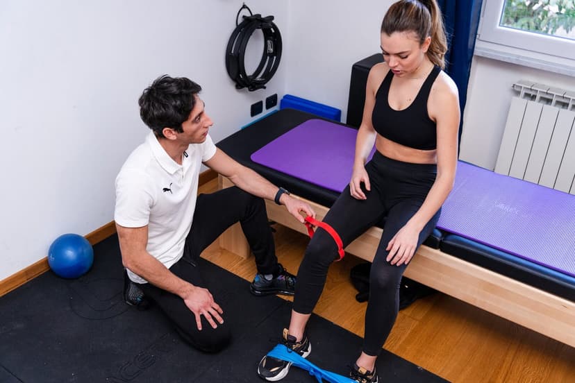
Hip work. Glute bridges, side-lying abductions, band exercises above the knees. This is the part the consensus recommends alongside the knee and the one most often skipped in practice.
Extensions and progressive loads. When the symptoms allow, the knee should be brought back to tolerate higher loads, otherwise the improvement stalls halfway. This is the phase in which having someone watch your technique makes the difference.
The exact doses, how many repetitions, how many seconds, how many times a week, vary enormously from one study to another, and nobody has shown that one formula is superior to the others. Consistency over weeks and the fact that the load really rises over time matter far more.
Stretching, mobility and stiffness
Stretching the quadriceps, the hamstrings, the calves and the outer hip has always been in the toolbox, and it is reasonable when you find obvious stiffness that limits movement.
It should, however, be placed in the right spot: in consensus documents therapeutic exercise is the main intervention, while stretching and soft-tissue techniques remain complementary. If you have half an hour, spend most of it loading the knee progressively, not pulling the iliotibial band with a foam roller.
The other treatments: what the consensus recommends
Here the literature is clearer than one might expect.
- Insoles. The 2018 consensus recommends them to improve pain and function, and the 2024 guide lists them among the supportive interventions, in the ready-made version. They are an aid, not a structural correction.
- Taping and kneecap braces. They are classified among the interventions on which there is uncertainty: they may help some people in the short term, but there is no consensus on their effect. The idea that they "recentre" the kneecap should be taken with a pinch of salt.
- Isolated mobilisations of the kneecap, the knee or the lumbar region, and electrophysical agents (the various passive instrumental therapies): not recommended by the 2018 consensus.
- Acupuncture and dry needling, soft-tissue manual techniques, blood flow restriction training, gait retraining: a zone of uncertainty, to be evaluated case by case.
- Ice and anti-inflammatories can make an acute phase manageable. They are tools to let you do the rest, not treatments in themselves, and medicines should be used for short periods and on medical advice.
- Injections and surgery. The options exist (arthroscopic debridement, realignment procedures, up to patellofemoral replacement in extreme cases), but they remain a last resort: as a review devoted precisely to patellar chondromalacia notes, none of the approaches proposed for cartilage lesions in this area has established itself as a standard, either for symptoms or for preventing the progression to osteoarthritis.
How long it takes, said honestly
There is no guaranteed time to get out of patellar chondromalacia, and anyone who promises you one is guessing.
What we know is that this pain is not as self-limiting as was believed: in the follow-up study cited above, more than half of the people traced at 5 to 8 years reported an unfavourable recovery, a category that, as said, also includes those who had improved moderately. The same authors identified two factors that at the time of diagnosis predict a worse course: symptoms lasting more than 12 months and a poorer function score at the start. And they conclude with a recommendation addressed to colleagues before patients: educate people to have realistic expectations.
Two practical indications follow. The first: do not wait months hoping it will pass, because the time you let slip by is itself a prognostic factor. The second: if after weeks of work done properly nothing moves, the answer is not to push the same thing harder, it is to reassess.
Prevention and load management
In practice, preventing patellar chondromalacia coincides with common sense applied to load.
Increase training volumes gradually, giving the knee time to adapt. Do not return to activity after a long break by picking up where you left off. Vary the stimuli instead of repeating the same identical movement for months. Wear shoes suited to the activity you do and replace them when they are visibly worn. Maintain general fitness: a trained knee tolerates more.
And above all, do not treat the knee as a fragile object. Fear of movement is a factor that has been measured in this condition: a systematic review of 41 articles and 2,712 people with patellofemoral pain found a moderate association between higher levels of kinesiophobia and poorer function as reported by the patient (n = 499, r = -0.440), and a weak association with pain intensity (n = 644, r = 0.162). It is a statistical link, not proof of cause, but it is enough to justify the authors' recommendation to take it into account in assessment, and to suggest that frightening someone with the word "wear" is not a neutral act.
TeleRehabilitation as an option
The thread that runs through all the evidence in this article is that the value lies in a progressive programme followed over time, supported by a clear explanation of what is happening. And that is exactly what gets lost when you download a sheet and carry it out alone for two weeks.
With Ri-Hub a physiotherapist assesses your situation, sets the load on the basis of what you can tolerate today and updates it session after session, correcting the execution on video call. For a condition in which consistency matters more than the choice of the individual exercise, being followed from home removes precisely the friction that makes people give up.
You might also be interested in:
Anterior Knee Pain: Causes, Diagnosis and Treatment Starting to Exercise: A Complete Guide for Beginners Plantar Fasciitis: Immediate Remedies for the Pain
Stay in touch with us Join our dedicated Facebook group: we answer all patients, give practical advice and clear up the most common doubts.
Sources
- Prevalence of knee osteoarthritis features on magnetic resonance imaging in asymptomatic uninjured adults: a systematic review and meta-analysis, British Journal of Sports Medicine, 2019
- Exercise for treating patellofemoral pain syndrome, Cochrane Database of Systematic Reviews, 2015
- 2018 Consensus statement on exercise therapy and physical interventions (orthoses, taping and manual therapy) to treat patellofemoral pain, British Journal of Sports Medicine, 2018
- Best practice guide for patellofemoral pain based on synthesis of a systematic review, the patient voice and expert clinical reasoning, British Journal of Sports Medicine, 2024
- Factors that predict a poor outcome 5-8 years after the diagnosis of patellofemoral pain: a multicentre observational analysis, British Journal of Sports Medicine, 2016
- Effectiveness of Isolated Hip Exercise, Knee Exercise, or Free Physical Activity for Patellofemoral Pain: A Randomized Controlled Trial, The American Journal of Sports Medicine, 2019
- Does atrophy of vastus medialis obliquus and vastus lateralis exist in patients with patellofemoral pain syndrome, Journal of Orthopaedic Surgery and Research, 2021
- 2016 Patellofemoral pain consensus statement from the 4th International Patellofemoral Pain Research Retreat, Manchester. Part 1, British Journal of Sports Medicine, 2016
- Chondromalacia patellae: current options and emerging cell therapies, Stem Cell Research & Therapy, 2021
- Kinesiophobia Is Associated With Poor Function and Modifiable Through Interventions in People With Patellofemoral Pain: A Systematic Review With Individual Participant Data Correlation Meta-Analysis, Physical Therapy, 2023
- Knee crepitus is prevalent in women with patellofemoral pain, but is not related with function, physical activity and pain, Physical Therapy in Sport, 2018
Last source check: August 2026.
Need a consultation?
Start the Ri-Hub questionnaire: a few questions about your case, you sign up, and you see who will follow you: a physiotherapist on the Italian professional register, whom you can write to in chat whenever you like. If you would rather talk it through by voice, the first consultation is free, lasts twenty minutes and commits you to nothing: they arrive having already read what you wrote, and together you decide whether and how to go on.


